Monday, March 30, 2015


Welcome back to Dylan and Devin Discovering Dirt (D^4). This week we preformed a stain that Devin and I have been looking forward to for a few weeks now, the Endospore Stain.


Before we get into our experiment there are some questions that need to be answered.
  1. What are endospores?
  2. What are the benefits of endospores?
  3. Why might some microbes have evolved to form endospores?
  4. Why might some microbes have evolved to NOT form endospores?
1. Well, according to sciencedaily.com:
  • Endospore-  a dormant, tough, non-reproductive structure produced by a small number of bacteria from the Firmicute family.
These endospores are formed in times of environmental stress such as changes in temperature or lack of nutrients. Bacterial form endospores as a way of bunkering down until environmental conditions get better. Think of endospore production like Birmingham in a snow storm- Production (Cell growth) within the city essentially shuts down, and everyone takes shelter (Endospore) until the snow has melted, at which point production (Cell growth) continues.
http://www.sciencedaily.com/articles/e/endospore.h
Above the process of endospore formation can be see.
Figure credit: http://academic.pgcc.edu/~kroberts/Lecture/Chapter%203/03-22_Sporulation_L.jpg


2. Endospores can be advantageous for bacterial cells. As seen above, endospore formation results in a DNA surrounded by a cell membrane and a tough spore coat. This spore coat results in resistance to heat (>100C), radiation, acids, bases, alcohol, and desiccation. As you could imagine, the previously mentioned environmental factors are account for a great number of cell deaths, so having a mechanism for combating these environmental changes can be very advantageous. Microbiologytext.com even reports that endospores from 25-40 million year old amber have been germinated in medium.
You can read more about that, and more on endospores here:
http://www.microbiologytext.com/index.php?module=book&func=displayarticle&art_id=69


3. The trait of endospore formation is undoubtedly a shining example of evolution. Due to the beneficial nature of endospore formation, it is feasible that an obligate bacteria that evolved this trait was a lone survivor of a change in environment at which point the trait became successful. Endospore formation would allow for the survival of bacteria that would otherwise be selective in the environments in which they could persist.


4. In contrast, some bacteria are less sensitive to changes in environmental changes, in which case the formation of a vegetative endospore could be a disadvantage. Furthermore, there are instances in which an environmental change will persist for a short period of time, then subside. In this case, it may be more advantageous to remain in a non-endospore state and utilize resources that are now available due to the vegetation of endospore-forming bacteria. In another way of thinking, the "Endospore trait" may not be seen in all bacteria because their are other methods of resisting environmental stress.


Now that we know a bit more about Endospores lets dive into this weeks experiment.


Devin and I preformed an Endospore Stain as well as an Endospore Growth Assay! An endospore stain results in endospores showing up as a green, and the cytoplasm of the cell as pink.


Condensed Procedure:
Endospore Staining
  1. Prepare a smear of unknown, Endospore positive, Endospore negative bacteria on respective slides.
  2. Flood slides with malachite green, while suspending the slides over a steaming hot beaker of water.
  3. Rinse with water.
  4. Counterstain with aqueous safranin
  5. Examine under microscope.
Results:


Example of successful Endospore stain of Endospore forming bacteria. Photo Credit: http://academic.missouriwestern.edu/jcbaker/bio251sec01/DSC02875.JPG
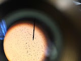
Results of endospore staining our unknown soil microbe. Taken in lab March 24, 2015.
       
As can be seen above our bacteria has a rather peculiar appearance. This week we obtained a stain with the least amount of contamination of any stain we have attempted. Interestingly, we since preforming the gram stain, we were predicting our bacteria was an endospore former, due to it's pleomorphic shapes, and unusual white circles in every cell. To our surprise the results from this endospore stain suggest our bacteria is actually not and endospore forming bacteria. This can be seen by the lack of green color in the above stain. For further assurance of the status of our bacteria we also preformed an Endospore Growth Assay.


Endospore Growth Assay

  1. Six 2ml tubes of tryptic soy broth were inoculated with one 3 different bacteria.
    1. Bacillus (Positive control)
    2. E.coli (Negative control)
    3. Our unknown microbe)
  2. Each bacteria were separated into two treatments:
    1. Heat Shock
    2. Non-Heat Shock
  3. Heat-Shock bacteria were incubated in a 80C heat bath for 10 minutes.
  4. After heat shocking all treatments were left to sit for 4 days to measure growth.
Results.


Above the results of the Endospore Growth Assay can be seen. From left to right the treatments are: Positive control (Bacillus ....), Heat Shock Bacillus, Negative control E.coli, Heat shocked E.coli,, unknown control, unknown Heat shock.


As seen in the gloriously blurry picture above (adding pictures to BlogSpot requires a master of witchcraft. Seriously.) Our positive control, a species of Bacillus showed precipitate in both the control and Heat shock trials (as expected for an endospore former). In excellent micro-lab fashion, our negative control E.coli also showed precipitate across both the control and heat shock trials. This was unexpected as E.coli is a known non endospore former. There must have been a failure in our technique via either contamination with an endospore former, or we were unsuccessful in killing the E.coli in the heat shock sample. Even more intriguing is the results of our unknown bacteria. Our bacteria showed precipitate in both the control and the heat shock treatments. This leads to further confusion as to whether our methodology was compromised, and to whether our bacteria is actually an Endospore Former.


The Dirty Truth About Our Microbe!
As Devin discussed last week, we now know quite a bit about our soil microbe


"Thus far we have conducted five experiments to further our knowledge of our unknown soil microbe. We have done a Gram stain to determine if our microbe is gram positive or gram negative, we have tested to see if our microbe was acid or non-acid fast, we have tested for catalase activity, we have tested to see if our microbe is aerobic or aerobic, and now we have determined if our microbe is an endospore former.

          We have determined so far, that our bacteria is neither gram positive or gram negative, but rather a bacteria called endospore-forming bacteria, which is a mix of bacilli and cocci shaped bacteria. We have also determined that our bacteria is non-acid fast due to the color it appeared after staining. There was also no sign of bubble formation during the catalase test which means that our bacteria has no catalase activity. Finally, we tested to see if the bacteria was an aerobic or anaerobic bacteria, and the results stated that there was activity of both types of metabolism. "

After this week we are once again left to interpret our results lightly. We found that our stain showed our microbe in the best clarity of any stain thus far, but it did not suggest we are working with an endospore former. Conversely our Endospore Growth Assay provided evidence that our microbe does a dandy job of surviving high temperatures through endospore formation. Since our results thus far have been a bit inconclusive in a few areas; I set out on a quest to the vast expanses of the internet to gain some more knowledge on our pleomorphic bacteria.

My quest for dirty knowledge was fruitful (Thanks Dr. Hanson) as there may be a potential order of bacteria that fits with our unknown microbe: Actinomycetales. More specifically the genus Streptomycetes. Members of the Actinomycetes Order:
  • form branching filaments of cells which become a network of strands called a mycelium; this week we noticed thin filamentous projections protruding from our bacteria.
  • Produce Spores in a Unique way.
  • Gram positive
Our mystery microbe aligns with these characteristics in these ways nearly identically. (Depending on your interpretation of some of our results.)


Streptomyces spores
Streptomyces bacteria. Photo credit:
 http://www.microbiologybytes.com/video/Streptomyces.html

Above is the first image of our bacteria obtained during our Gram-Stain.


In comparing the picture of Streptomyces to our unknown microbe taken on day one, there is a strong argument to be made that there is a similarity. Both display similar pleomorphic shapes, with a mysterious round white center.
Further investigations need to be done to confirm this information, but I believe we are on the right track. Streptomyces are reported as having no flagella, so next week we will preform a motility stain which will help in supporting or ending our case for the unknown soil microbe being Streptomyces.
Join us next time as Devin Breaks it down and discusses Motility and our Flagella Stain! Is our mystery microbe flagellin?


Monday, March 23, 2015

CATALASE ACTIVITY
For our sixth lab Dylan and I tested our unknown microbe to see if it had any catalase activity. What is a catalase you may ask? A catalase is an enzyme that is found in the majority of living organisms that are exposed to oxygen. The enzyme catalyzes the decomposition reaction of hydrogen peroxide into the separate elements, water and oxygen.

This photo was taken from the Birmingham-Southern 
Microbiology Lab Manual for Week Six of BI 304    
In order to determine if our unknown had catalase activity or not, a very small amount of our microbe was placed on a sterile microscope slide. Once the microbe sample was on the slide one drop of 3% H2O2  was dropped onto the sample, and then the slide was sealed in a petri dish to eliminate contamination of aerosols. If the slide had immediate formation of bubbles then the result of the catalase test was positive, but if there was no bubble formation then the test was negative. This reaction can be seen in the picture to the right.
This test is done to differentiate catalase-positive Micrococcaceae from catalase-negative Streptococcaceae. Upon testing our microbe it was observed that there was no formation of bubbles, which means it was a negative catalase test or a catalase-negative Streptococcaceae. Additional characteristics that can be determined from a catalase activity test are determining if a bacteria is gram-positive or gram-negative and differentiating between aerobic and obligate anaerobic bacteria. 
The reason that some microbes have evolved to have catalase activity is because of how harmful hydrogen peroxide can be when produced by metabolic processes. In order for the organisms that produce hydrogen peroxide as a byproduct of these metabolic processes to be safe from its effects they catalyze the compound into safer substances (water and oxygen). Many organisms must rely on “defense mechanisms that allow them to repair or escape the oxidative damages of H2O2" in order to survive.

CARBOHYDRATE METABOLISM
             The next step in determining the species of our unknown soil microbe was to run a Triple Sugar Iron Test. A TSI Test is a tube of slanted agar medium that is used in order to determine if the production of hydrogen sulfide is present and to see is there is fermentation of carbohydrates. Three different substances can be used in order to differentiate fermentation and those are, sucrose, lactose, and glucose. TSI tests are also very useful for determining different types of gram‐negative bacteria.  
             The tube to the right shows what a TSI slant looks like before a sample has been placed on the medium. The red color of the medium comes from the pH indicator (phenol red). The slant is made up of an aerobic (oxygen) section which is placed near the top of the tube, while the bottom of the tube, also known as the butt, is anaerobic (no oxygen).
             Once the microbe sample has been placed on the slant a variety of results can appear; 1.)Glucose Fermenters, 2.)Glucose, Sucrose, and Lactose Non-fermenters 3.) Glucose, Sucrose and/or Lactose Fermenters 4.)Glucose Fermenter and Hydrogen Sulfide Producer, and 5.)Glucose, Lactose and/or Sucrose Fermenter and Hydrogen Sulfide Producer. There could also be there two cases of Gas Producers and Glucose Non‐fermenter Hydrogen Sulfide Producers. The pictures below show what the TSI slants would look like if any of the five results listed about were to happen.


Far Left: Glucose Fermenter
Second from Left:Glucose, Sucrose and/or Lactose       Fermenter
Second from Right:Glucose Fermenter and Hydrogen Sulfide Producer
Far Right:Glucose, Lactose and/or Sucrose Fermenter and Hydrogen Sulfide Producer.
Not Pictured: Glucose, Sucrose, and Lactose Non-fermenters


             The controls used for the TSI portion of the experiment were E. coli (A/A), B. megaterium (A/NC), P. areginosa (K/K) and P. vulgaris (A/A + H2S). A/A stands for acid over acid, which means that glucose, lactose and/or sucrose have been metabolized. A/NC stands for   acid over no change. K/K stands for alkaline over alkaline, which indicates that all three of the sugars have been metabolized. A/A + H2S stands for acid over acid with the production of a black precipitate at the butt of the tube. 
           The following pictures show the four control slants and then our unknown microbe slant after the butt had been punctured and the slant had been spread across the media.

Left: P. vulgaris (A/A + H2S), middle: P. areginosa (K/K), and right: B. megaterium (A/A).
Left: our unknown (?/?) and right: E. coli (A/A).
           Did our slants react in the way we expected them to? For the majority yes, except for P. vulgaris. P. vulgaris is (A/A + H2S) which means that it should've turned yellow in the early hours of the experiment, and then result in a sold black substance at the butt of the tube. This black substance that was supposed to be seen is the precipitate of the hydrogen peroxide, but it was not observed when the slant was checked on after incubation. As for the rest of the slants they all showed expected results. The B. megaterium metabolized glucose very quickly in the beginning stage of the experiment, which kept it the red color of the slant; ammonia was then released in the anaerobic butt of the slant which caused the medium to turn yellow. P. areginosa also followed the expected results by going from pink to red, which indicates that all three of the sugars were metabolized. 
         For our unknown the color of the media near the top of the tube was a yellow-orange color, and so was the butt of the slant. It was also determined, after observation, that our unknown has no evidence of hydrogen sulfide formation due to the lack of black substance in the butt. So we have come to the conclusion that our unknown must be very similar to. E. coli. They looked very similar after incubation, which leads us to believe that our unknown is also a Glucose, Lactose and/or Sucrose Fermenter just like E.coli. They both produced expected results by turning a yellow-orange color. This change symbolizes the fast metabolism of glucose, lactose and/or sucrose. 

SLOWLY DISCOVERING OUR DIRT 
         Thus far we have conducted five experiments to further our knowledge of our unknown soil microbe. We have done a Gram stain to determine if our microbe is gram positive or gram negative, we have tested to see if our microbe was acid or non-acid fast, we have tested for catalase activity, and most recently have tested to see if our microbe is aerobic or aerobic.
          We have determined so far, that our bacteria is neither gram positive or gram negative, but rather a bacteria called endospore-forming bacteria, which is a mix of bacilli and cocci shaped bacteria. We have also determined that our bacteria is non-acid fast due to the color it appeared after staining. There was also no sign of bubble formation during the catalase test which means that our bacteria has no catalase activity. Finally, we tested to see if the bacteria was an aerobic or anaerobic bacteria, and the results stated that there was activity of both types of metabolism. 

STAYED TUNED NEXT WEEK FOR DYLAN'S NEXT INSTALLMENT OF D^4 WHEN WE DIG FURTHER INTO FIGURING OUT THE CLASSIFICATION OF OUR UNKNOWN MICROBE!!!

Tuesday, March 10, 2015

Welcome to Week Four of Dylan and Devin Discovering Dirt.


As you remember, last week Devin and I preformed a Gram-Stain, and grew a culture of our unknown microbe on a Maconkey Auger. The results from these trials gave us evidence that our bacteria is a Gram-Positive bacteria. This means our microbe has a dense Peptidoglycan cell wall!


To further understand the makeup of our soil microbe's cell wall, this week we preformed an "Acid-Fast" Stain. The purpose of this stain is to determine if the cultured cells exhibit high lipid content cell walls (characteristic of all acid-fast bacteria). Specifically, the lipid in question is Mycolic Acid, which causes the cell walls of acid-fast bacteria to be waxy and very selective permeability. 


Identifying this trait is helpful in identifying bacteria, because only a few genera of bacteria possess the acidfast property, Nocardia and Mycobacterium. This trait is also great for identifying many medically important bacteria, such as Mycobacterium tuberculosis, the causative agent of tuberculosis.


So, with that lets discuss how the Ziehl-Neelsen Method of acid-fast stain works.


Why Acid Stain?

  • Acid-fast bacteria have a waxy and nearly impermeable cell wall, due to the aforementioned mycolic acid.
  •  Water based stains (such as the Gram stain) are unable to adequately penetrate this waxy mycolic acid.
  • The Waxy Layer must be removed by heat to stain the cell.


Methodology-


  1. Create a smear using a drop of water and a dab of cultured microbe.
  2. Place slides over beaker of steaming water for 1 minute.
  3. While over the beaker, flood the slide with Carbol-Fuchsin, and allow to sit over steam for 5 minutes.
  4. Allow the slide to cool after 5 minutes and rinse with tap water.
  5. Add acid alcohol to the slide until color no longer runs from the smear.
  6. Rinse with tap water.
  7. Flood with methylene blue and allow to react for 1 minute.
  8. Rinse with tap water and blot dry.
  9. Examine with microscope.
The Dirty Science-

  • The Primary stain (pink) is driven into acid-fast cells during the heating period
  • When cooled, Acid-Alcohol is applied to remove Carbol fuchsin from cells without mycolic acid.
  • Crystal violet counterstain infiltrates bacterial cell walls that do not contain mycolic acid. (similar to a gram stain)
  • Acid Fast= Pink  
  •  Non-Acid-Fast= Purple
An example of ideal results of an Acid-fast Stain.
Photo: http://imc02.hccs.edu/BiologyLabs/Micro/01MicrobialMorphologyStaining/01BacterialStaining.html
Dirty Results
As it can be seen in the picture above, our results actually found a grey area between acid-fast and non-acid-fast. When compared to the ideal examples of acid-fast and non-acid-fast stains, our unknown microbe seems to be a perfect fit in-between both colors. (Maybe it's like #thedress an it's really white and gold?) We can not say with confidence whether our bacteria is acid-fast or not, but to me it seems the cell walls of the bacteria are a bit closer to blue than pink, so tentatively we will say our bacteria is non-acid-fast until further experiments are conducted.


With the results we have collected so far we can begin to attempt to use a dichotomous key to narrow down the possible identities of our microbe.


Dichotomous Key results:
  1. Gram Positive
  2. Bacilli
  3. Non Acid-Fast
  4. Catalase positive or negative?


The dichotomous key leads us to the next characteristic we need to determine: is our microbe a Catalase producer? Catalase is an enzyme some bacteria secrete as a defense against Hydrogen Peroxide. Until we determine this characteristic of our microbe, we have only ruled out the Genus Mycobacterium and Norcardia. This will be our focus in our lab experiments in the coming week.


Tune in next time as we preform our catalase and carbohydrate test!


 You Stay Classy San Diego


Information on the science of Acid-Fast Staining was obtained at the following site:


http://www.scienceprofonline.com/microbiology/acid-fast-ziehel-neelsen-bacteria-stain-identify-mycobacteria-nocardia.html

Monday, March 2, 2015

Hello Everyone!
      Welcome to Week Three of Dylan and Devin Discovering Dirt! This past week Dylan and I made another leap and bound into our soil microbe research. The objective of the last step in our research was to determine if the soil microbe that we have specifically chosen is Gram-positive or Gram-negative. What are Gram-positive and Gram-negative cells you ask? Let me give you the DIRTY details.
      First, you need to know what a Gram stain is. A Gram stain is the method of taking bacteria and separating them into two distinct groups by using the physical and chemical properties of their cell walls to determine if peptidoglycan is present. Peptidoglycan is a polymer made of amino acids and sugars. These amino acids and sugar combine to form a net-like layer on the outside of the plasma membrane is the majority of bacteria, and this membrane is known as a cell wall. The amount of peptidoglycan in the cell wall is what differentiates Gram-positive from Gram-negative. 

This figure shows the difference in the amount of peptidoglycan present in a Gram-positive cell wall in comparison to a Gram-negative cell wall. It can be seen that the peptidoglycan layers in the cell wall of the Gram-positive bacteria are MUCH thicker than the single layer in the the Gram-negative bacteria.
Gram-positive Bacteria
      Now for the DIRTY details of what differentiates a Gram-positive and Gram-negative bacteria. Gram-positive bacteria are bacteria that give positive results in a Gram stain test. The bacteria absorb the crystal violet stain used in the test, which results in the purple appearance seen when looking at the bacteria through the microscope. This purple color is seen because the thick peptidoglycan layer in the bacterial cell wall has the ability to retain the crystal violet stain after it is washed away from the rest of the sample, in the decolorization stage of the test. 


Gram-negative Bacteria 
      Gram-negtive bacteria are unable to retain the crystal violet stain after the decolorization step. The alcohol used during decolorization degrades the outer membrane of gram-negative bacteria, which makes the cell wall more porous resulting in the inability to retain the crystal violet stain. The peptidoglycan layer of Gram-negative bacteria is much thinner and is in between an inner cell membrane and a bacterial outer membrane. This placement causes them to take up the counterstain (safranin or fuchsine), which causes the bacteria to appear pink.

The Gram staining process.
      So, now that you know the differences between Gram-positive and Gram negative bacteria and the process of a Gram stain, it's time to see how Dylan and I did in staining a bacteria for ourselves! Dylan and I went through the process of smearing our unknown bacteria onto a microscope slide. We then proceeded to flood the smear with crystal violet dye. Once the dye had been on the smear for a minute its was rinsed off with water, and then Gram's iodine was added to the smear to soak for one minute. After the Gram's iodine sat for one minute it was rinsed off gradually with 95% ethanol to remove any excess crystal violet dye on the smear. Once all excess dye was removed the Gram's safranin counterstain was added to the smear for 30 seconds and then rinsed off. It can be seen in the picture of the Gram stain process the Dylan and I stained our unknown bacteria (bottom slide: UK), a Gram-negative bacteria (middle slide: K. pneumonia), and a Gram-positive bacteria (top slide: B. megaterium).
     
     
Unknown bacteria slide through the
microscope lens.
      After the Gram staining process and after looking at our unknown slide in comparison to our Gram-positive and Gram-negative control slides under the microscope Dylan and I were unable to make a decision in full confidence as to which type of bacteria our unknown was. It can be seen in the picture to the left that out bacteria is purple in color, which lead us to come to the conclusion that our sample was Gram-positive but we were still uncertain. The uncertainty is due to the fact that the shapes of our bacteria aren't consistent throughout the entire smear. 
Coccus                                                         Bacillus                                                  Diplobacillus 
      The three types of bacterial shapes above are the bacterial shapes Dylan and I believe are seen in our unknown bacterial sample. After doing a little bit our research and consulting with our lab instructor Dylan and I arrived at the conclusion that our unknown sample was a type of bacteria known as endospore-producing bacteria. An endospore-producing bacteria is also known as sporulating bacteria. Sporulating bacteria are derived from the phylum Firmicute and consist mostly of Gram-positive bacteria . These endospore-forming bacteria belong primarily to the Bacillus and Clostridium genus. So, even though Dylan and I didn't get our prediction 100% accurate, we did come close in saying that the unknown was endospore-producing because endospore-producing bacteria are primarily Gram-positive. 

MacConkey agar plate including the unknown
bacteria, B. megaterium (+), K. pneumonia (-),
P. aurigenosa (-).
      Aside from looking at the color and the shape of the unknown bacteria through the microscope and comparing them to the Gram-positive and Gram-negative control bacteria, Dylan and I also set up a MacConkey agar in order to observe Gram-positive and Gram-negative growth. MacConkey agar is used for the isolation of Gram-negative enteric bacteria and the differentiation of lactose fermenting from lactose non-fermenting Gram-negative bacteria. A compact version of the T-streak method was used on the MacConkey in order to test all four bacteria on one plate. When looking at the plate, it can be seen that the two Gram-negative controls both showed growth, but the Gram-positive control and the unknown bacteria had no growth. The fact that the B. megaterium and the unknown bacteria did not have any growth occur is just more evidence to prove that Dylan and I were correct in predicting that our unknown bacterial sample is Gram-positive. 

     To wrap up the blog Dylan and I have decided to do some critical thinking and answer the question, "How does the Gram status influence the treatment of bacterial infections?" Gram-positive bacteria have a much greater amount of peptidoglycan in their cell membrane, which means that they have a very thick outer layer of peptidoglycan around their cell wall. This thick outer layer, has the ability to absorb large amounts of foreign material. Gram-negative bacteria, on the other hand, have a very thin outer membrane. Even though the membrane of Gram-negative bacteria is very thin it is very difficult to penetrate. Because of their thin but strong cell membrane, gram-negative bacteria are often resistant to many types of antibiotics. The level of difficulty associated with penetrating the cell wall of a Gram-negative bacteria is one of the reasons that Gram-negative bacteria are harder to treat than Gram-positive bacteria.
      Another reason, and probably the most important reason, that Gram-negative bacteria are harder to treat than Gram-positive is their resistance to drugs. Dr. Brad Spellberg, an infectious-disease specialist at Harbor-U.C.L.A. Medical Center in Torrance, Calif., and the author of “Rising Plague,” a book about drug-resistant pathogens states that, “For Gram-positives we need better drugs; for Gram-negatives we need any drugs.” Dr. Azza Eleman also said, “You don’t really have much choice, if a person has a life-threatening infection, you have to take a risk of causing damage to the kidney.” This quote is in reference to the only two drugs really used for treating a Gram-negative bacterial infection; collision and polymyxin B. These drugs were established in the 1940's and have really not been put to much use since then because they have such deleterious effects on the kidney and cause serious nerve damage. Although these drugs have horrible side effects, like Dr. Eleman said, they have been very rarely used which means that the Gram-negative bacteria have not built up a resistance to them.
      So in conclusion, Gram-positive bacterial infections are much easier to treat than Gram-negative bacterial infections because even though they have a thicker peptidoglycan layer they do not possess an outer membrane. The lack of an outer membrane in Gram-positive bacteria allows their membranes to be penetrated much more easily than Gram-negative bacteria. This means that aside from very high drug resistance causing treatment difficulty, Gram-negative bacteria is also difficult to treat because it contains the outer membrane surrounding its thin peptidoglycan layer.

http://www.nytimes.com/2010/02/27/business/27germ.html?_r=0

JOIN US NEXT WEEK TO SEE WHAT DYLAN HAS TO SAY ABOUT ACID-FAST BACTERIA!!!